What is a Hybrid Prosthesis in Implant Dentistry?

Implant dentistry can really change people's lives.  In cases where there have been significant amounts of bone and tissue loss, this type of restoration allows the patient to regain both the function and the look of a real dentition. Here is a rough outline of the process:

1.  Before the treatment was executed, proper diagnostics were done with study models, X-rays, CT Scan, etc.

2.  The implants were then placed, and allowed to integrate with the patient’s bone.

3.  An accurate impression was done to capture the implant positions and was sent to the laboratory.

4.  The lab then created a duplicate model of the patient’s arch (with the implants replicated exactly how they are in the mouth).

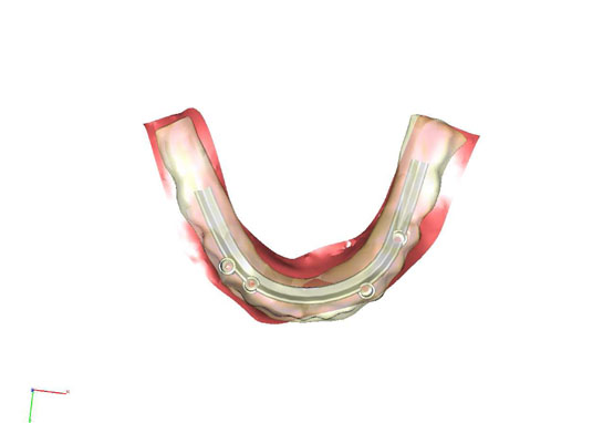
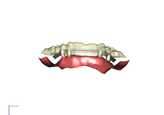
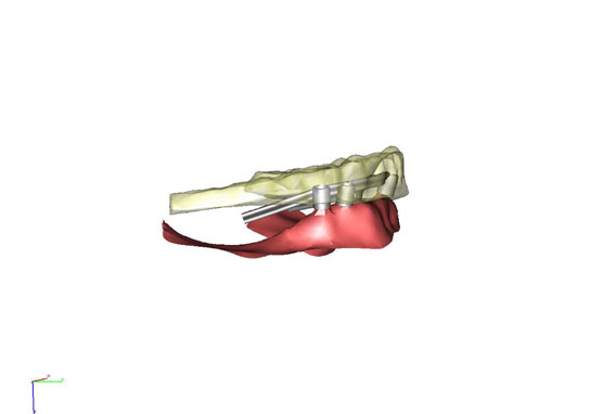
5.    A Cad Cam scan was done to design a titanium framework for the prosthesis.  See my blog entry dated Dec 4, 2010 about the Cad Cam technology for this same case.

6.  The titanium bar is tried in the mouth on the actual implants to ensure accurate fit.

7.  Teeth and gum colored materials are placed over the titanium bar.

8.  The final prosthesis was placed precisely over the implants in the patient’s mouth.  The entire prosthesis was held in placed by screws that fit into the implants.  Openings at the screw access were filled with proper composite materials to blend in with the surrounding areas.

9.  Proper contacts and functional guidance of the bite were checked and fine-tuned.

The patient once again has a full arch of teeth fixated to his/her arch to replace the missing teeth.  This type of restoration takes away the pain and inefficient chewing problem for patients who maybe suffering from the lack of ability to simply … eat.

Case Photos:

Images of actual patients of Alex Nguyen, DDS are Copyrighted and Digitally Embedded to track Unauthorized Use.

The titanium bar on the stone model:

The prosthesis on the stone model.  Note the access holes where the implants were positioned.

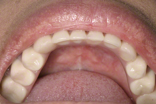
The final hybrid prosthesis in the mouth.  Note all implant access holes were filled.

The patient's smile at completion of treatment.

........................................................................................

Alex Nguyen, DDS is a Saratoga Dentist who practices General Dentistry, Cosmetic, and Implant Dentistry. For over 20 years the practice has been serving the residents of Santa Clara County and San Francisco Bay Area.

CAD/CAM Technology in Dental Implant Treatments

CAD/CAM is a technology utilized in many aspects of dentistry. In dental implant treatment for patients who have lost all teeth, we can take advantage of this powerful tool to survey our patient's dentition, review existing position of implants, and design the final prosthesis. Today's sophisticated computer softwares allow the dentist and lab team to review restorative cases in multiple dimensions before fabricating the prosthesis.

In the example below we were able to identify a positioning problem with one of the implants.  Rather than producing a compromised restoration, we were able to resolve the issues before proceeding.

This is one of the many ways we deliver quality practice to our patients everyday.

........................................................................................

Alex Nguyen, DDS is a Saratoga Dentist who practices General Dentistry, Cosmetic, and Implant Dentistry. For over 20 years the practice has been serving the residents of Santa Clara County and San Francisco Bay Area.

Problems with Root Canal Teeth and the Importance of Proper Advanced Planning for Dental Implants

Root Canals will alleviate pain from the affected teeth.  However, when root canals are not done properly problems may not be evident until much later. This patient described the symptom as "some discomfort around my old crown".

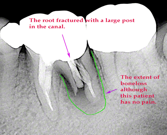
Thorough clinical exam and xray revealed a fractured root where a large post was placed in the canal.  The root canal and crown were done many years ago. Root canal treated teeth have a higher chance of root fracture. Although the patient had no pain after the treatment, the failed root canal and post placement sustained a low grade infection around the root area. Where there are infections, bacteria slowly cause the jaw bone to break down.

In areas where the bone is extremely dense, the patient may not have any symptom until too much bone have been lost.  In the xray below we can see the outline of the extent of the  bone loss.  What you cannot see when looking at the area inside the mouth is an entire wall of bone that has been destroyed.  That wall of bone used to exist between the root and the gum tissue on the out side.

So why is this discussion important in treating these kinds of problems?

If we simply extract this tooth, the bone loss area will collapse.  After the area has healed, any subsequent grafting procedures and treatment planning for dental implant in the area may become extremely difficult to carry out.

If your treatment preference is to replace this tooth with a dental implant, it is crucial that you consult with the right clinician to properly treatment plan the situation.  Carrying out the treatment with proper advanced planning will minimize further complications to the area.

Images of actual patients of Alex Nguyen, DDS are Copyrighted and Digitally Embedded to track Unauthorized Use.

........................................................................................

Alex Nguyen, DDS is a Saratoga Dentist who practices General Dentistry, Cosmetic, and Implant Dentistry. For over 20 years the practice has been serving the residents of Santa Clara County and San Francisco Bay Area.
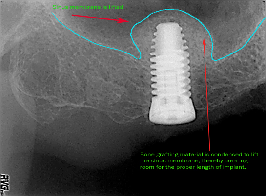
What is a Sinus Lift for Dental Implant?

Sometimes there is not enough bone in the areas of the upper molars and bicuspids because the sinus floor has a tendency to expand into the space after the teeth have been lost.  In order to properly anchor the right implant length for strength, we need to recreate adequate bone height. With today's innovation in instrumentation and technique, we can predictably condense bone grafting material and lift the floor of the sinus to create room for the implant.  After the healing period, the bone solidifies around the implant and the result is much more predictable than to use a short or mini implant.

Images of actual patients of Alex Nguyen, DDS are Copyrighted and Digitally Embedded to track Unauthorized Use.

........................................................................................

Alex Nguyen, DDS is a Saratoga Dentist who practices General Dentistry, Cosmetic, and Implant Dentistry. For over 20 years the practice has been serving the residents of Santa Clara County and San Francisco Bay Area.

A Second Opinion on Dental Treatments - The Importance of the Quality and Type of Xray!

This past week a patient came to see me for a second opinion on her dental implant treatment.  She brought me a copy of the xray below, and asked me if I could advise her on whether or not she should do 1 or 2 implants to replace the teeth indicated by the green arrows.  She wanted to find out which of the following treatment options is better:

Note:  After this xray was taken, tooth (10) was extracted and a bone graft was done for this area by an oral surgeon.  In other words, tooth (10) currently is missing.

Plan A:  Extract the teeth labeled (8) and (9); place an implant at (8); do a bridge from position (8) to (11).

Plan B:  Keep teeth (8) and (9) as is, and simply attach a new tooth to the implant crown (11) to replace missing tooth (10).

Unfortunately, I could not answer her questions because the copy of the xray given to her was not useable.  Printed on regular white printer paper, the xray was dark and fuzzy. I could not clearly see the bone level at the location of these teeth. In fact, to identify the health of the existing teeth, other types of xrays would be necessary to have for our evaluation of the current condition.  In this case, xrays called periapicals (often abbreviated PA) would then allow us to not only evaluate the bone but also the root structure and internal condition of these teeth. For example, if the integrity of teeth (8) and (9) is normal, and a successful bone graft was done for (10),  then why would we want to remove 2 additional fully functional teeth?

We simply did not have enough information to determine an accurate evaluation based on this xray alone.  Furthermore, when it comes to implant treatment, accurate studies of this patient's bite also would be necessary in order to complete a diagnosis and treatment plan.

When asking for copies of your xrays, please remember to ask your dentist to provide you with quality copies. This most basic diagnostic tool must be clear and readable.